Summary
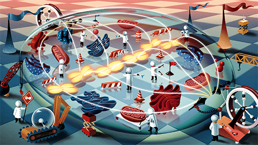
 As the mitotic spindle tightens to separate chromosomes during cell division, it produces and absorbs forces.
Ada Zejun Shen/Quanta Magazine
Introduction
The cells of animals, plants, and fungi start their lives by being torn apart. Cells are born by division, and just before a parent cell becomes two daughters, it doubles its nuclear DNA and carefully condenses it into X-shaped chromosomes. The nucleus disassembles, letting these crucial genetic instructions float free in the cell’s soupy interior. Then the cell performs an astounding, microscopic feat of strength.
Proteinaceous cables extend from the cell’s poles toward the equator and latch onto the chromosomes. They drag, tilt, and nudge the precious cargo until every chromosome has been ushered into a tidy line around the cell’s middle. Then this spindle apparatus, as it’s known — a sinewy, dynamic rib cage made of bundles of microtubules — shortens itself at both poles. This wrenches the chromosomes apart into two sets and reels them to opposite ends of the cytoplasm sea. With its genetic material segregated at either pole, one cell can safely become two, born from a microscopic tug-of-war.
The spindle strains against itself as it shortens and pulls; how it does this without ripping itself apart has been a scientific mystery since biophysicists first observed cell division with microscopes 150 years ago. “They saw them [the chromosomes] moving, which led to this idea that there’s probably forces that are pulling or pushing things around,” said Colleen Caldwell, a biophysicist at the University of Groningen.
If absorbing those forces caused the spindle’s integrity to fail, it could spell the end for both daughter cells or cause diseases that arise from errors in cell division and chromosome arrangement. In this way, all eukaryotic life, including human life, rides on the spindle’s success with each cell division across an organism’s lifetime.
Until recently, researchers didn’t have the tools to physically manipulate the mammalian spindle structure at the subcellular scale to toy with it and find out how it works. Recently a team of researchers led by Sophie Dumont, a biophysicist at the University of California, San Francisco, used microneedles to physically manipulate and stress the structure in mammal cells for the first time — and then observe how the spindle holds together through intense strain as it wrenches the chromosomes apart.
The experiments have shown how a self-repair mechanism enables the spindle to stabilize itself under force and avoid disintegrating. These findings, which were published in February 2026 in Current Biology, provide a window into the physics of the cellular world, where complex living machines endure physical forces and stresses like machines in a factory. The spindle’s mechanical quirks show just how weird materials science can get at the finest scales of life.
A Living Material
By virtue of being biological, the cell spindle presents massive complexity for materials physicists. Most human-made materials contain just a few different types of molecules, said Colm Kelleher, a biophysicist at Syracuse University who was not involved with the new research. Meanwhile, the spindle is made of hundreds of different types of individual protein molecules, and any one of them is “an extremely complex object,” he said.
That puts the spindle in an unusual size class that complicates experiments. “There’s quite a bit that scientists know about the mechanics of individual molecules, and there’s quite a bit that scientists know about the mechanics of tissues and organisms, like how muscles generate force,” Dumont said. “But mechanics at this scale of many molecules together forming this macromolecular structure is harder to probe. So we know less about it, but it’s just as important.”
One last wrinkle is that, by being part of a living organism, these biomolecular structures are constantly consuming energy from within the materials themselves — very unlike how human-made materials and machines work. Kelleher gave the example of a car: It has a fuel tank and an engine, which power components that transfer torque to the wheels, which then push against the ground. A system made of biological materials works very differently.
Mark Belan/Quanta Magazine
“It would be like if you had a car where there were only wheels, and you injected the gasoline directly into the wheel, and it all started moving itself,” he said. “The force-generating components, the energy-consuming components, and the force-transferring components are all physically mixed up with each other.”
Despite the mechanical oddities inherent to living machines, investigations into spindle physics have been going on for decades. In the 1960s, the late Duke University biologist Bruce Niklas started using extremely fine glass needles to probe and manipulate chromosomes in living cells by pushing from the outside against the cell membrane. By exerting physical force on the spindle and chromosomes, he and his colleagues revealed some of the key mechanics for the first time. For example, tension on the kinetochores — the disc-shaped proteins to which the spindle’s fibers attach on the chromosome — is thought to let the cell confirm the spindle’s correct attachment and ensure the proper separation of chromosomes during cell division.
As the mitotic spindle tightens to separate chromosomes during cell division, it produces and absorbs forces.
Ada Zejun Shen/Quanta Magazine
Introduction
The cells of animals, plants, and fungi start their lives by being torn apart. Cells are born by division, and just before a parent cell becomes two daughters, it doubles its nuclear DNA and carefully condenses it into X-shaped chromosomes. The nucleus disassembles, letting these crucial genetic instructions float free in the cell’s soupy interior. Then the cell performs an astounding, microscopic feat of strength.
Proteinaceous cables extend from the cell’s poles toward the equator and latch onto the chromosomes. They drag, tilt, and nudge the precious cargo until every chromosome has been ushered into a tidy line around the cell’s middle. Then this spindle apparatus, as it’s known — a sinewy, dynamic rib cage made of bundles of microtubules — shortens itself at both poles. This wrenches the chromosomes apart into two sets and reels them to opposite ends of the cytoplasm sea. With its genetic material segregated at either pole, one cell can safely become two, born from a microscopic tug-of-war.
The spindle strains against itself as it shortens and pulls; how it does this without ripping itself apart has been a scientific mystery since biophysicists first observed cell division with microscopes 150 years ago. “They saw them [the chromosomes] moving, which led to this idea that there’s probably forces that are pulling or pushing things around,” said Colleen Caldwell, a biophysicist at the University of Groningen.
If absorbing those forces caused the spindle’s integrity to fail, it could spell the end for both daughter cells or cause diseases that arise from errors in cell division and chromosome arrangement. In this way, all eukaryotic life, including human life, rides on the spindle’s success with each cell division across an organism’s lifetime.
Until recently, researchers didn’t have the tools to physically manipulate the mammalian spindle structure at the subcellular scale to toy with it and find out how it works. Recently a team of researchers led by Sophie Dumont, a biophysicist at the University of California, San Francisco, used microneedles to physically manipulate and stress the structure in mammal cells for the first time — and then observe how the spindle holds together through intense strain as it wrenches the chromosomes apart.
The experiments have shown how a self-repair mechanism enables the spindle to stabilize itself under force and avoid disintegrating. These findings, which were published in February 2026 in Current Biology, provide a window into the physics of the cellular world, where complex living machines endure physical forces and stresses like machines in a factory. The spindle’s mechanical quirks show just how weird materials science can get at the finest scales of life.
A Living Material
By virtue of being biological, the cell spindle presents massive complexity for materials physicists. Most human-made materials contain just a few different types of molecules, said Colm Kelleher, a biophysicist at Syracuse University who was not involved with the new research. Meanwhile, the spindle is made of hundreds of different types of individual protein molecules, and any one of them is “an extremely complex object,” he said.
That puts the spindle in an unusual size class that complicates experiments. “There’s quite a bit that scientists know about the mechanics of individual molecules, and there’s quite a bit that scientists know about the mechanics of tissues and organisms, like how muscles generate force,” Dumont said. “But mechanics at this scale of many molecules together forming this macromolecular structure is harder to probe. So we know less about it, but it’s just as important.”
One last wrinkle is that, by being part of a living organism, these biomolecular structures are constantly consuming energy from within the materials themselves — very unlike how human-made materials and machines work. Kelleher gave the example of a car: It has a fuel tank and an engine, which power components that transfer torque to the wheels, which then push against the ground. A system made of biological materials works very differently.
Mark Belan/Quanta Magazine
“It would be like if you had a car where there were only wheels, and you injected the gasoline directly into the wheel, and it all started moving itself,” he said. “The force-generating components, the energy-consuming components, and the force-transferring components are all physically mixed up with each other.”
Despite the mechanical oddities inherent to living machines, investigations into spindle physics have been going on for decades. In the 1960s, the late Duke University biologist Bruce Niklas started using extremely fine glass needles to probe and manipulate chromosomes in living cells by pushing from the outside against the cell membrane. By exerting physical force on the spindle and chromosomes, he and his colleagues revealed some of the key mechanics for the first time. For example, tension on the kinetochores — the disc-shaped proteins to which the spindle’s fibers attach on the chromosome — is thought to let the cell confirm the spindle’s correct attachment and ensure the proper separation of chromosomes during cell division.
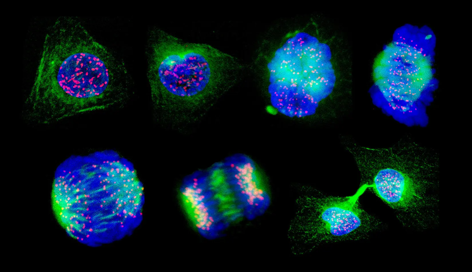
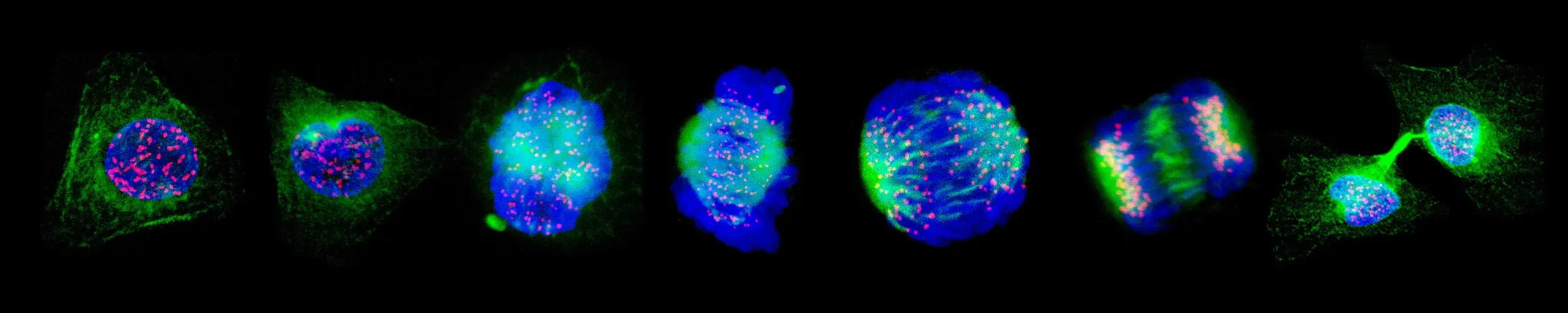 The stages of cell division. The cell spindle (green) attaches to the chromosomes at their kinetochores (pink). It lines them up in the middle of the cell and then pulls them apart to opposite poles. Then the cell splits into two.
Wellcome Images/Science Source
Over the next few decades, Niklas’ work prodding and pulling on the fibers started the field of spindle mechanics, Dumont said. He is “basically our hero,” she added. His work opened up questions about how the spindle generates force, how much force it can produce, and how it responds to any forces the environment exerts on it.
However, his work was done in a very specific type of cell: grasshopper spermatocytes, the progenitors of grasshopper sperm cells. These cells had some experimental benefits. They tolerate physical manipulation with glass microneedles, for instance, and have large, easily observable chromosomes. But Dumont wanted to look beyond insects to find out know how the spindle behaves in mammalian cells like ours. She needed to find a type of mammal cell that, like Niklas’ grasshopper spermatocytes, had large but relatively few chromosomes and were amenable to manipulation by microneedles.
The cells of rat kangaroos, rabbit-size nocturnal marsupials, have only 12 or 13 chromosomes depending on the sex. They turned out to be ideal. For approximately 10 years, Dumont’s lab has tugged on rat kangaroo cells with microneedles — “sometimes we call it cell torture,” she said — to see how they respond to different forces.
A Spindle’s Resilience
In 2020, her team got a surprise. When they yanked on the fiber with the microneedle, it did not detach from the chromosome or from the spindle’s anchor points at the cell’s poles. The fiber broke in the middle, like a pencil snapping in two. What’s more, the ragged ends of the busted fibers didn’t immediately unravel. They settled into a stable form, like a nylon rope whose frayed end has been melted to stop further disintegration.
Caleb Rux, a bioengineer in the Dumont lab, looked more closely at how the fibers were breaking and somehow being stabilized afterward. To manipulate the spindle with extreme precision, he used what looked like a video game controller to lower a motorized microneedle and move it in any direction in 62.5-nanometer increments.
“I think of it as like a 3D Etch a Sketch,” Rux said. The faster the needles were programmed to move and pull against the spindle fibers, the more force they applied.
Rux, Dumont, and their colleagues found that when fibers were cut with lasers, which don’t exert a tugging force, they became unstable and fell apart. But fibers that were first stressed and stretched with a microneedle and then cut in that spot with a laser were able to stabilize.
Dumont’s team wondered whether there was a kind of automatic repair mechanism that could heal spans of the fiber when it was subjected to force. The spindle fibers are microtubules, which are essentially long rods made up of many tiny interlocking components. Rux compared those components to Lego bricks that assemble into a much larger structure. “Under force, these individual Lego [bricks] might pop out of the structure — not at the end, but literally in the middle of it,” Rux said. And when they do, more stable bricks in the area could pop in to take their place and stabilize the fiber.
The stages of cell division. The cell spindle (green) attaches to the chromosomes at their kinetochores (pink). It lines them up in the middle of the cell and then pulls them apart to opposite poles. Then the cell splits into two.
Wellcome Images/Science Source
Over the next few decades, Niklas’ work prodding and pulling on the fibers started the field of spindle mechanics, Dumont said. He is “basically our hero,” she added. His work opened up questions about how the spindle generates force, how much force it can produce, and how it responds to any forces the environment exerts on it.
However, his work was done in a very specific type of cell: grasshopper spermatocytes, the progenitors of grasshopper sperm cells. These cells had some experimental benefits. They tolerate physical manipulation with glass microneedles, for instance, and have large, easily observable chromosomes. But Dumont wanted to look beyond insects to find out know how the spindle behaves in mammalian cells like ours. She needed to find a type of mammal cell that, like Niklas’ grasshopper spermatocytes, had large but relatively few chromosomes and were amenable to manipulation by microneedles.
The cells of rat kangaroos, rabbit-size nocturnal marsupials, have only 12 or 13 chromosomes depending on the sex. They turned out to be ideal. For approximately 10 years, Dumont’s lab has tugged on rat kangaroo cells with microneedles — “sometimes we call it cell torture,” she said — to see how they respond to different forces.
A Spindle’s Resilience
In 2020, her team got a surprise. When they yanked on the fiber with the microneedle, it did not detach from the chromosome or from the spindle’s anchor points at the cell’s poles. The fiber broke in the middle, like a pencil snapping in two. What’s more, the ragged ends of the busted fibers didn’t immediately unravel. They settled into a stable form, like a nylon rope whose frayed end has been melted to stop further disintegration.
Caleb Rux, a bioengineer in the Dumont lab, looked more closely at how the fibers were breaking and somehow being stabilized afterward. To manipulate the spindle with extreme precision, he used what looked like a video game controller to lower a motorized microneedle and move it in any direction in 62.5-nanometer increments.
“I think of it as like a 3D Etch a Sketch,” Rux said. The faster the needles were programmed to move and pull against the spindle fibers, the more force they applied.
Rux, Dumont, and their colleagues found that when fibers were cut with lasers, which don’t exert a tugging force, they became unstable and fell apart. But fibers that were first stressed and stretched with a microneedle and then cut in that spot with a laser were able to stabilize.
Dumont’s team wondered whether there was a kind of automatic repair mechanism that could heal spans of the fiber when it was subjected to force. The spindle fibers are microtubules, which are essentially long rods made up of many tiny interlocking components. Rux compared those components to Lego bricks that assemble into a much larger structure. “Under force, these individual Lego [bricks] might pop out of the structure — not at the end, but literally in the middle of it,” Rux said. And when they do, more stable bricks in the area could pop in to take their place and stabilize the fiber.
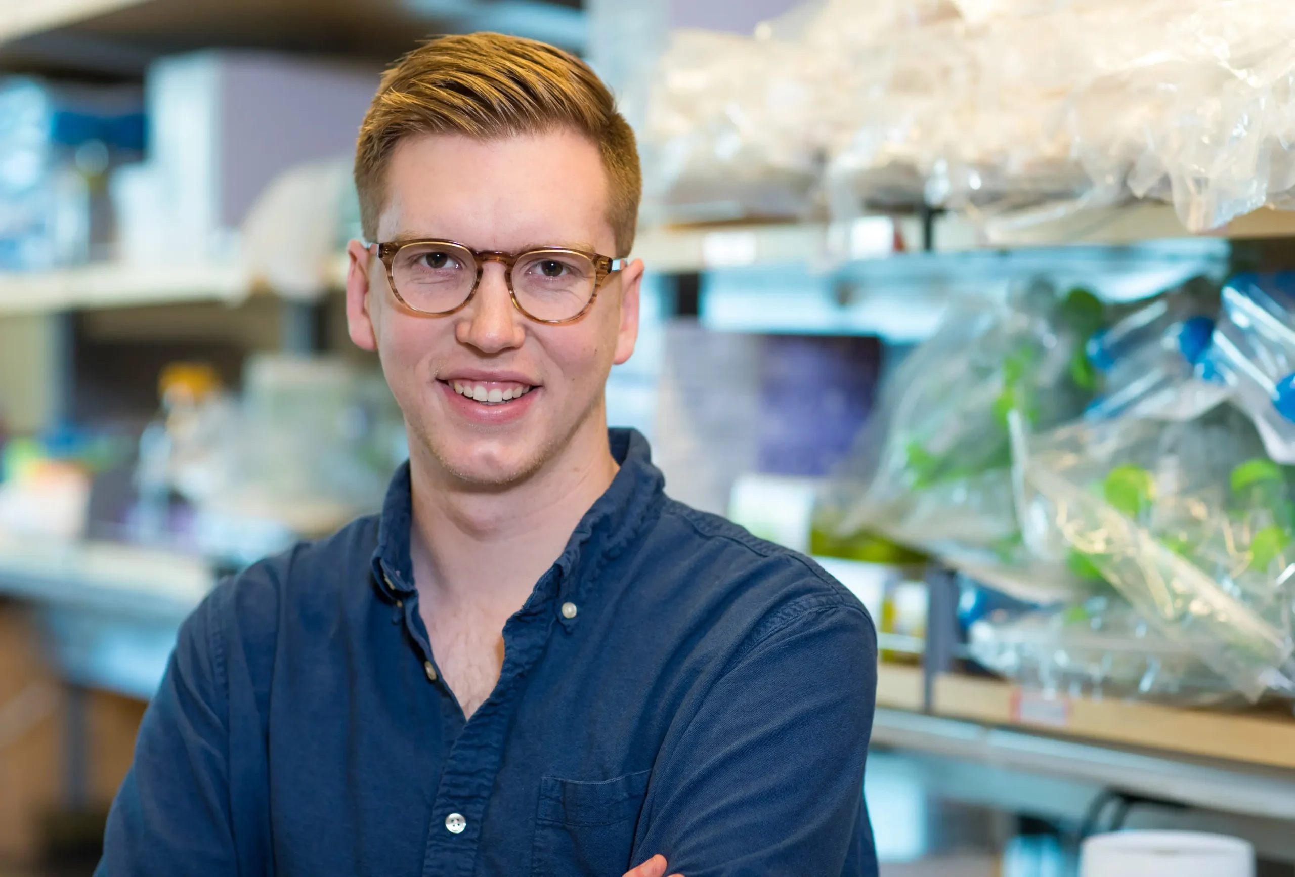 Caleb Rux led work that revealed the resilience of the mitotic spindle, the critical structure that organizes DNA during cell division. “It took years to get to that mechanism,” he said.
Elisabeth Fall
To test their hypothesis, they needed to know where such repairs might be happening. They added a fluorescent tag to end-binding protein 1 (EB1), a microtubule-associated protein that prefers to bind to stable compounds rather than less stable ones. The fiber lit up with this protein at the exact same spot where force was being exerted, suggesting that self-stabilization was occurring.
“It took years to get to that mechanism,” Rux said. “It was really exciting and made me feel vindicated that all the reading and work we’d done had actually led us down the right road.”
He hypothesizes that when force is applied to the spindle fiber, the stretching and pulling makes it jettison some of its smaller cytoskeletal components — the Lego bricks — which get pushed out into the surrounding cytoplasm. At the same time, other, more stable bricks suspended in the cytoplasm can pop into the injured structures and reinforce them. This makes the entire spindle structure more stable precisely where it needs support. This may be an automatic process, Rux said, or specific proteins might encourage the microtubule bricks to fill the gaps.
The phenomenon is a bit counterintuitive. Alexander Mogilner, a computational biologist at New York University who was not involved in the study, compared it to a finger trap. Normally when you pull on something, it breaks. But here, pulling makes the material stronger. “The brute message of the study is kind of heartwarming — that the external forces would stabilize the spindle and make it more resilient,” he said.
This mechanism may explain how the spindle manages to exert and withstand the forces generated when it plays chromosomal tug-of-war. The spindle’s life, after all, is a bit of a paradox, Dumont said.
“It has to be a dynamic structure to build itself, to move chromosomes, to remodel, yet it has to be strong,” she said. Perhaps a workable solution is to spontaneously gird and stabilize itself when generating and absorbing force.
The lesson of spindle resilience extends beyond the biological. Rux used to work as a mechanical engineer inspecting roads for cracks that result from use and weathering. He now wonders if it’d be possible to engineer roads that grow stronger under load, rather than more fragile over time.
“Biology’s had billions of years to evolve these structures to be really robust and good at their jobs,” he said. “I think there are things to be learned from some of these principles, and [we can] use [them] to inspire some of our engineered systems as well.”
Caleb Rux led work that revealed the resilience of the mitotic spindle, the critical structure that organizes DNA during cell division. “It took years to get to that mechanism,” he said.
Elisabeth Fall
To test their hypothesis, they needed to know where such repairs might be happening. They added a fluorescent tag to end-binding protein 1 (EB1), a microtubule-associated protein that prefers to bind to stable compounds rather than less stable ones. The fiber lit up with this protein at the exact same spot where force was being exerted, suggesting that self-stabilization was occurring.
“It took years to get to that mechanism,” Rux said. “It was really exciting and made me feel vindicated that all the reading and work we’d done had actually led us down the right road.”
He hypothesizes that when force is applied to the spindle fiber, the stretching and pulling makes it jettison some of its smaller cytoskeletal components — the Lego bricks — which get pushed out into the surrounding cytoplasm. At the same time, other, more stable bricks suspended in the cytoplasm can pop into the injured structures and reinforce them. This makes the entire spindle structure more stable precisely where it needs support. This may be an automatic process, Rux said, or specific proteins might encourage the microtubule bricks to fill the gaps.
The phenomenon is a bit counterintuitive. Alexander Mogilner, a computational biologist at New York University who was not involved in the study, compared it to a finger trap. Normally when you pull on something, it breaks. But here, pulling makes the material stronger. “The brute message of the study is kind of heartwarming — that the external forces would stabilize the spindle and make it more resilient,” he said.
This mechanism may explain how the spindle manages to exert and withstand the forces generated when it plays chromosomal tug-of-war. The spindle’s life, after all, is a bit of a paradox, Dumont said.
“It has to be a dynamic structure to build itself, to move chromosomes, to remodel, yet it has to be strong,” she said. Perhaps a workable solution is to spontaneously gird and stabilize itself when generating and absorbing force.
The lesson of spindle resilience extends beyond the biological. Rux used to work as a mechanical engineer inspecting roads for cracks that result from use and weathering. He now wonders if it’d be possible to engineer roads that grow stronger under load, rather than more fragile over time.
“Biology’s had billions of years to evolve these structures to be really robust and good at their jobs,” he said. “I think there are things to be learned from some of these principles, and [we can] use [them] to inspire some of our engineered systems as well.”